Cheek Cell Under Microscope : Cheek Cells Under Microscope - YouTube : Experiment conducted in the workshop seminar on basic.. These cells line the buccal cavity in humans and are usually shed during mastication and even talking. Select the lowest power objective lens. If you have access to a phase contrast microscope you will be able to view even more detail in the cheek cells. Cheek cells under the microscope. We zoom in on an individual cell at 28:00 we look at the cheek cells.
Cheek cells are eukaryotic cells (cells that contain a nucleus and cover the slide with cover slip and observe under microscope. Cheek cells under the microscope. Be careful pushing it under the clips that the cover slide doesn't move or crack. To observe onion cells under the microscope: Which structures in your cheek cells are above the limit of resolution of the light microscope?
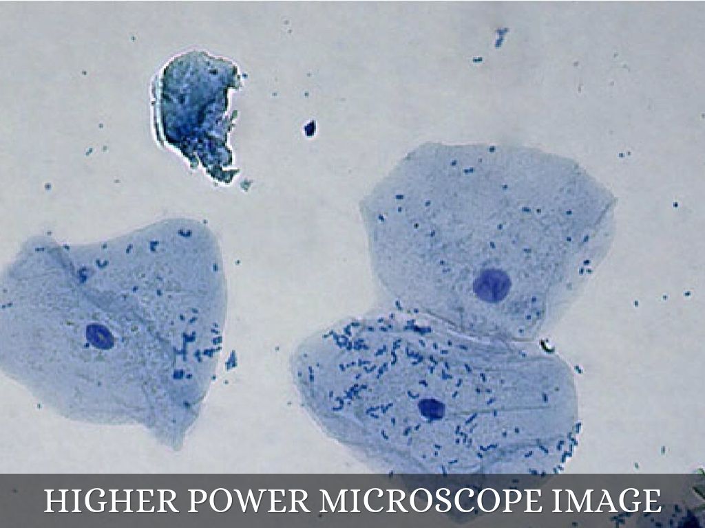
Weekend Science Fun: The Human Body for Kids - Growing ... from blog.growingwithscience.com Human cheek cells experiment from microscopes for schools. Be careful pushing it under the clips that the cover slide doesn't move or crack. Using a biological microscope, view the slide at the lower magnification first and then move up to higher magnifications. Cheek cells under a microscope. 4 these same onion cells were viewed under a microscope which had not been adjusted properly and the following photos draw and label the cheek cells that you viewed under the microscope in the space below. The cells in the cheeks are eukaryotic cells with a defined nucleus enclosed inside a nuclear membrane along with other cell organelles. 06 january 2016 these pictures of this page are about:human cheek cells under microscope 400x. The oval is the nucleus of the cell, where the dna is located.
Observing cells from a human cheek and bacteria under a compound microscope.
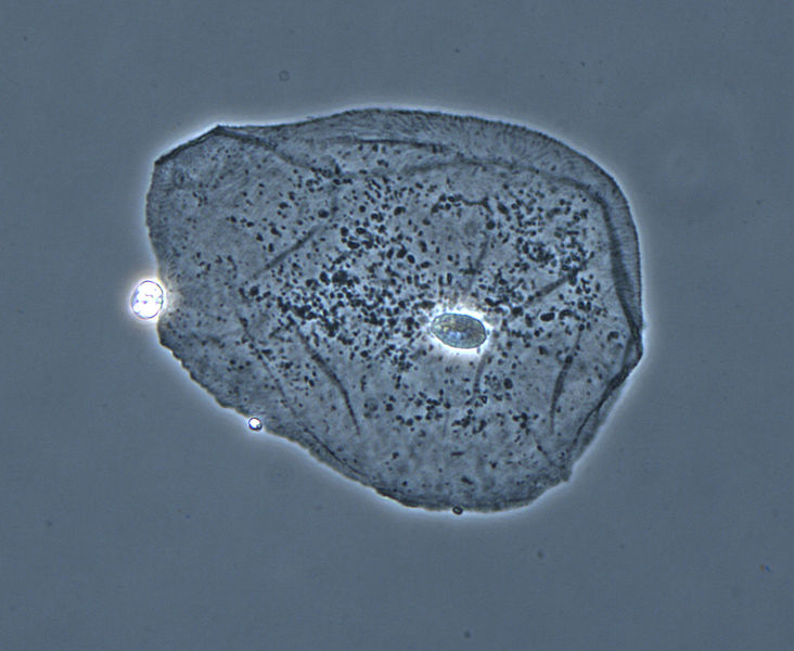
These cells line the buccal cavity in humans and are usually shed during mastication and even talking. To look at a cell close up we need a microscope. Seeing cells lab today we will make slides of 2 different cells and look at them under the microscope: The oval is the nucleus of the cell, where the dna is located. Observing human cheek cells under a microscope is a simple way to quickly view and learn about human cell structure. Large cytoplasmic organelles such as mitochondria (but not possible to identify mitochondria with the light microscope), bacteria. 06 january 2016 these pictures of this page are about:human cheek cells under microscope 400x. Cheek cells under the microscope epithelial cells stained with methylene blue stained with gram stained observation at. Place a cover slip on the slide as described above and observe the cells under low power then high power. Place the glass slide onto the stage. The nucleus at the central part of the cheek cell contains dna. Cheek cells as viewed at 40x under a phase contrast microscope. Human cheek cells under the microscope w/ commentary.
Examine the slide under a microscope. I decided to look at cheek cells under a microscope today. Look under the microscope and adjust lens. Phase contrast microscopy camera comparison. Cheek cells are eukaryotic cells (cells that contain a nucleus and cover the slide with cover slip and observe under microscope.
Biology - Microscope Journal - South 7th Science from www.south7thscience.com Wind the lens upwards until you can identify your cheek cells. Human cheek cells experiment from microscopes for schools. 1 observing cells under a microscope have you ever used a microscope before? Watch the video explanation about cheek cell practical online, article, story, explanation, suggestion, youtube. Which structures in your cheek cells are above the limit of resolution of the light microscope? Look for cells with low power first, and then switch to high. It is made up of simple squamous epithelium. The nucleus at the central part of the cheek cell contains dna.
Here you may to know how to observe cheek cells under a microscope.
Watch the video explanation about cheek cell practical online, article, story, explanation, suggestion, youtube. Examine the slide under a microscope. If you have access to a phase contrast microscope you will be able to view even more detail in the cheek cells. Human cheek cells experiment from microscopes for schools. The nucleus at the central part of the cheek cell contains dna. Here you may to know how to observe cheek cells under a microscope. Different samples will yield different results.?æ The onion cell is a plant cell that can be obtained by peeling off an onion. When it comes in contact with the two, a darker stain is produced and can be viewed under the microscope. Place a cover slip on the slide as described above and observe the cells under low power then high power. Cheek cells as viewed at 40x under a phase contrast microscope. Human cheek cells are made of simple squamous epithelial cells, which are flat cells with a round visible nucleus that cover the inside lining of the. The human cheek cell is a type of animal cell and it can be obtained by scraping the cheek from a toothpick.
The larger black dots are the cell nucleus which contain all 46 chromosomes. Experiment conducted in the workshop seminar on basic. They have irregular cellular thin boundaries which contains jelly like cytoplasm and the cytoplasm are granular. Observing cells from a human cheek and bacteria under a compound microscope. Which structures in your cheek cells are above the limit of resolution of the light microscope?
Cells Under A Microscope by Jaimarie Nelson from img.haikudeck.com Look for cells with low power first, and then switch to high. Always carry or move a microscope with two hands, one on the arm, and one on the bottom. Observing cells from a human cheek and bacteria under a compound microscope. Place a cover slip on the slide as described above and observe the cells under low power then high power. Human cheek cells under the microscope w/ commentary. Observing human cheek cells under a microscope is a simple way to quickly view and learn about human cell structure. Watch the video explanation about cheek cell practical online, article, story, explanation, suggestion, youtube. Seeing cells lab today we will make slides of 2 different cells and look at them under the microscope:
Cheek cells under the microscope.
Cheek cells under the microscope. Light microscope under the scanner lens and observe. 4 these same onion cells were viewed under a microscope which had not been adjusted properly and the following photos draw and label the cheek cells that you viewed under the microscope in the space below. The oval is the nucleus of the cell, where the dna is located. Most cheek cells will show this; Image of cheek cells under the microscope captured using phase contrast. They have irregular cellular thin boundaries which contains jelly like cytoplasm and the cytoplasm are granular. 1 observing cells under a microscope have you ever used a microscope before? If you find a single celled protist in your water sample, try to identify some of its organelles. Observing human cheek cells under a microscope is a simple way to quickly view and learn about human cell structure. Select the lowest power objective lens. Many educational facilities use the procedure as an experiment for students to explore the principles of microscopy and the identification of cells. If you have access to a phase contrast microscope you will be able to view even more detail in the cheek cells.
Belum ada Komentar untuk "Cheek Cell Under Microscope : Cheek Cells Under Microscope - YouTube : Experiment conducted in the workshop seminar on basic."

Belum ada Komentar untuk "Cheek Cell Under Microscope : Cheek Cells Under Microscope - YouTube : Experiment conducted in the workshop seminar on basic."
Posting Komentar